The viruses that infect bacteria are the most abundant biological entities on the planet. For example, a recent simple study of 92 showerheads and 36 toothbrushes from American bathrooms found more than 600 types of bacterial viruses, commonly called bacteriophages or phages. A teaspoon of coastal seawater has about 50 million phages.
While largely unnoticed, phages do not harm humans. On the contrary, these viruses are gaining increasing popularity as biomedicines to eradicate pathogenic bacteria, especially those associated with antibiotic-resistant infections.

READ MORE: Viruses are teeming on your toothbrush and shower head
READ MORE: Modified phage DNA can kill deadly pathogens
In a study published in the journal Nature Communications, Gino Cingolani, Ph.D., of the University of Alabama at Birmingham, and Federica Briani, Ph.D., of the Università degli Studi di Milano, Milan, Italy, have described the full molecular structure of the phage DEV. DEV infects and lyses Pseudomonas aeruginosa bacteria, an opportunistic pathogen in cystic fibrosis and other diseases. DEV is part of an experimental phage cocktail developed to eradicate P. aeruginosa infection in pre-clinical studies.
DNA pulled out of phage head
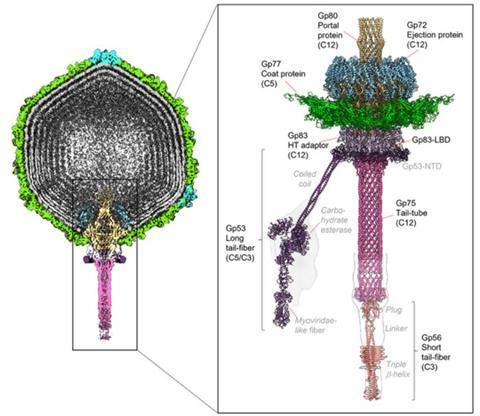
A peculiar feature of DEV is the presence of a 3,398-amino acid virion-associated RNA polymerase inside the capsid expelled into the bacterium upon infection. Unexpectedly, Cingolani and Briani’s study revealed the virion-associated RNA polymerase is part of a genome ejection motor that pulls the DNA of the phage out of its head after the phage has attached to the surface of a Pseudomonas bacteria using its tail fibers and has penetrated the cell’s outer and inner membranes using its tail tube.
“We posit that the design principles of the DEV ejection apparatus are conserved in all Schitoviridae phages,” Cingolani said. “As of October 2024, over 220 Schitoviridae genomes have been sequenced and are available in the public database. As these genomes are largely unannotated and many open-reading frames have unknown functions, our work paves the way for the facile identification of structural components when a new Schitoviridae phage is discovered.”
The Schitoviridae family of phages “represents some of biology’s most understudied bacterial viruses, increasingly utilized in phage therapy,” Cingolani said. “We are using structural biology to decipher the building blocks and map gene products. This is vital when the amino acid sequence evolves too rapidly for conventional phylogenetic analysis.”
Phage resemble lunar lander
The researchers used cryo-electron microscopy localized reconstruction, biochemical methods and genetic knockouts to describe the complete molecular architecture of DEV, whose DNA genome has 91 open-reading frames that include the giant virion-associated RNA polymerase. “This vRNAP is part of a three-gene operon conserved in all Schitoviridae genomes we analyzed,” Cingolani said. “We propose these three proteins are ejected into the host to form a genome ejection motor spanning the cell envelope.”
The structure of DEV and many other phages resembles a minuscule version of Neil Armstrong’s 1969 lunar lander, with a large head, or capsid, that contains the genome and leg-like fibers supporting the phage as it lands on the surface of bacteria, preparing to infect the living bacterial cell.

The researchers determined structures of all the protein capsid factors and tail components in DEV involved in host attachment. Through genetic experiments, they showed that the DEV long tail fibers were essential for infection of P. aeruginosa but were not needed to infect P. aeruginosa mutants whose surface lipopolysaccharide lacked the O-antigen. In general, viruses attach to different cell surface molecules as the first step of infection.
Stages of phage infection
While this study provides several still images of the phage structure, the researchers do not completely understand the movie of DEV infection. They envision three steps in that infection process.
In step one, as a single DEV phage drifts in isolation, its flexible long tail fibers fluctuate to improve the chance of touching a Pseudomonas lipopolysaccharide surface molecule. After the first touch, all five fibers attach to tether the phage perpendicularly close to the bacterial outer surface.
In step two, the short tail fiber, which also acts as a tail plug, touches a secondary receptor on the Pseudomonas and a mechanical signal releases the tail plug.
Up to this point, the three proteins called gp73, gp72 and gp71 have been stored inside the phage head near its tail, with shapes that will dramatically change when they exit the phage head. In step three, when the plug is gone, the three proteins are expelled out of the head and into the bacterial cell envelope.
The lead protein, gp73, refolds its shape to form an outer membrane pore with a hollow center. Below that, gp72 refolds into a hollow tube that spans the Pseudomonas periplasm, the space between the bacteria’s outer membrane and its inner membrane. Finally, gp71 crosses the inner membrane and refolds into a large RNA polymerase motor in the bacterial cytoplasm that pulls the phage DNA through the hollow gp73 and gp72 channels and into the Pseudomonas cell.


No comments yet