Antibiotics are widely used in clinical treatment and animal production as an effective means to combat microbial infections. Antimicrobial peptides, as potential alternatives to antibiotics, have shown promising applications.
However, owing to their broad‐spectrum antimicrobial activity, most antimicrobial peptides tend to cause imbalance in the host’s intestinal flora. In addition, the systemic use of broad‐spectrum drugs for the treatment of localized infections may cause toxicity, which should not be overlooked.
Phage display technology involves the fusion of DNA fragments encoding exogenous proteins or small‐molecule polypeptides with phage genes encoding surface proteins, which then exist on the surface of phages by means of fusion proteins. Exogenous proteins and small‐molecule polypeptides specifically recognize and bind to target molecules. Therefore, this technology is widely used to screen targeted drugs, prepare vaccines and monoclonal antibodies, and monitor and diagnose tumors.
Phage display technology
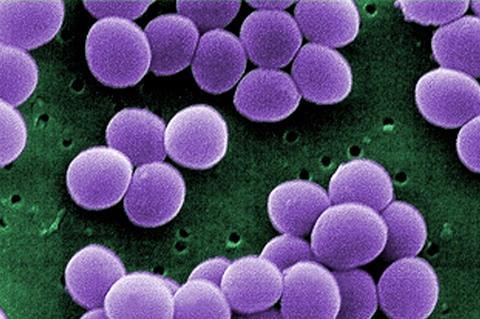
This study, published in mLife, screened nine heptapeptide sequences from a random peptide library using phage display technology, with S. aureus 6538 as the target bacterium. The heptapeptide sequence with the highest affinity (SYWVRAS) was selected as the target domain.
Researchers further improved its antimicrobial activity by adding several cationic amino acids (R or K) at the C‐terminus after linking the heptapeptide sequence to Temporin‐SHf, which is rich in hydrophobic amino acids, via GGG as a linker. Compared with Temporin‐SHf, the designed peptide had higher specific antimicrobial activity against S. aureus. This suggests that the screened heptapeptide sequence effectively improves the specific antimicrobial activity after concatenation with the template peptide.
Membrane integrity
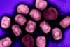
To further verify the effect of SFK2 treatment on membrane integrity, S. aureus 6538 was incubated with a final concentration of 8 μM SFK2, and the fluorescence distribution was observed under a laser scanning confocal microscope. Green fluorescence (SYTO9) and red fluorescence (PI) of S. aureus ATCC 6538 induced under 8 μM of SFK2 overlapped, indicating severe disruption of its cell membrane and leading to cell death.
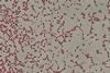
Subsequently, to investigate the targeting ability of SFK2, S. aureus ATCC 6538 and E. coli ATCC 25922 were mixed 1:1, treated with 8 μM SFK2, and then SYTO9 and PI fluorescent dyes were added for observation using laser scanning confocal microscopy. The green fluorescence and red fluorescence of S. aureus ATCC 6538 were induced to overlap at 8 μM concentration of SFK2, while E. coli ATCC 25922 only emitted green fluorescence without red fluorescence, indicating that its membrane integrity was not disrupted.
S. aureus, P. aeruginosa, and E. coli were incubated with different concentrations of SFK2, and their membrane integrity was quantified using flow cytometry. 8, 4, and 2 μM of SFK2 induced 92.78%, 70.08%, and 28.38% of S. aureus mortality, respectively. However, P. aeruginosa and E. coli maintained more than 90% survival even at the final concentration of 8 μM SFK2, with negligible differences from the control.
Morphological changes
To verify this hypothesis further, scanning electron microscope (SEM) and transmission electron microscopy (TEM) were used to observe the morphological changes in S. aureus treated with SFK2. The surface of untreated S. aureus was intact and smoothly rounded under SEM. S. aureus ruptured after 2 h of treatment with 2 μM SFK2, and there was an outflow of the contents outside the cell. Under the TEM, the cell membrane structure of S. aureus in the control group was complete, and the cytoplasm was dense. After treatment with 2 μM SFK2 for 2 h, the cell membrane of S. aureus was ruptured and the cell contents were discharged.
Researchers constructed and evaluated a mouse model of systemic infection using intraperitoneal. Both modes of administration significantly reduced the number of colonies in the organs, and symptoms such as hepatocellular rupture and inflammatory cell in-filtration were drastically reduced compared with those in the saline control. Because piglets are similar to humans in terms of anatomy, physiology, and nutritional metabolism, they further validated the in vivo effects of SFK2 using piglets as a model.
It was found that SFK2 remained active in vivo in a piglet model for treating S. aureus infection. These results indicate that the heptapeptide sequence screened using phage display technology can effectively improve the specific targeting ability of the peptide against S. aureus, providing a reference for the subsequent development of narrow‐spectrum antibacterial drugs.


No comments yet