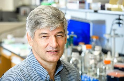
A $5.8 million grant led by Adrie Steyn, Ph.D., of the University of Alabama at Birmingham and the Africa Health Research Institute, or AHRI, in Durban, South Africa, will provide user-requested infected human lung tissue and analytical services to tuberculosis researchers worldwide.

Tuberculosis, or TB, is a bacterial infection that causes 1.3 million deaths and 10.6 million new active cases each year, yet experimental animal models of TB do not reproduce the full spectrum of disease as it occurs in humans. A paucity of human lung tissue for study has left a fundamental gap in understanding how the bacterial pathogen Mycobacterium tuberculosis, or Mtb, causes disease in people, says Steyn, a professor in the UAB Department of Microbiology.
READ MORE: Super cally molecules take down tuberculosis
READ MORE: Edited BCG offers potential vaccine to prevent tuberculosis in people of all ages
TB disease has existed since antiquity. Mtb was identified as the pathogen more than 140 years ago, and in 1800s Europe, TB caused nearly one in every four deaths. Human lung samples were highly studied until the disease was greatly diminished by better sanitation, and then the advent of antibiotics in the 1940s and ’50s. Since then, researchers have relied mainly on animal models to study TB.
Human pulmonary TB
Today, with the emergence of antibiotic-resistant Mtb, “there are few studies focused on elucidating in humans the fundamental mechanisms of active, subclinical and latent TB,” Steyn said. “We believe the TB field can no longer rely on animal models that yield findings of limited clinical relevance. Thus, there is an urgent need to better understand human pulmonary TB in order to develop clinically relevant therapeutic strategies.”
The five-year R24 grant from the National Institutes of Health will establish The Global Research Resource for Human Tuberculosis, with the goal of transforming the global landscape of TB research by accelerating the study of human TB tissue through two unique user-requested services. First is providing pathological, cellular, structural and genetic analysis of resected human TB or postmortem tissue. Second is disseminating human TB tissue to the global TB research community.
“We have several unique advantages since AHRI has access to a continuously growing collection of active TB/HIV and postmortem samples, including entire lungs and lobes,” Steyn said. “This R24 proposal is built upon substantial published data from our group that clearly demonstrate our capacity to examine resected and postmortem lung tissue from Mtb/HIV-infected human subjects. We consider the lack of human TB lung tissue to represent an unmet need and a critical barrier in the TB field that must be eliminated to develop new hypotheses.”
Survey for demand
To determine the need for human TB tissue and analytical services, Steyn and colleagues surveyed TB researchers through Mailchimp and X/Twitter, and nearly all respondents said they would highly value and use such a service.
In the first year of the grant, researchers will create repositories of human TB lung tissue for analysis and dissemination at AHRI, as well as existing pulmonary and extrapulmonary TB specimens at UAB and the University of Texas Halth Science Center, Houston. These will come from active, subclinical and latent TB patients and decedents, along with clinical and radiological information. About one-quarter of the world’s population is latently infected with Mtb, a poorly understood problem.
In the first year, the multi-center team will build a dedicated web portal to describe all its services, establish an international committee to rapidly evaluate applications through the web portal and establish a virtual information platform.
Over the next four years, the team will build service delivery and tissue distribution, with gradually increasing levels of service including increased volume or larger panels, flow cytometry of freshly resected TB tissue, laser capture microdissection, micro-computed tomography analysis of tissue for 3D reconstruction of the tuberculosis lung, fixed tissue sections or blocks, and whole-body X-ray imaging of decedents. In 2022, Steyn and colleagues were the first to use micro-computed tomography to reveal the three-dimensional microarchitecture of the human TB lung, and they showed that human TB granulomas are complex polymorphic shapes, not spherical structures.
Other analysis
Other analysis of human TB tissue will include immunohistochemistry and specialized stains, RNAscope to visualize individual mRNAs, spatial transcriptomics, and multiplex immunofluorescence. As part of their preliminary research, published in eBioMedicine this summer, Steyn and colleagues showed that the emerging technology, RNAscope, was able to identify Mtb mRNA in intact and disintegrating bacilli in tissues. They were able use RNAscope to identify Mtb mRNA in UAB lymph node biopsies taken more than a year before a patient died from disseminated tuberculosis. Those biopsies had been negative for the Ziehl-Neelsen stain, the standard test to identify Mtb. Steyn says this shows the diagnostic potential of RNAscope technology in complex TB cases, since the Ziehl-Neelsen stain is highly variable and can miss Mtb organisms.
AHRI is in an area with a high TB incidence and a large, well-characterized cohort of TB patients. Through decadeslong collaborations with cardiothoracic surgeons and anatomical and forensic pathologists at the Inkosi Albert Luthuli Central Hospital and the King DinuZulu Hospital, AHRI receives three or four freshly resected human lung tissue samples per week.
Clinically relevant research
Steyn says that, by providing the Global Research Resource for Human Tuberculosis service to the TB research community, many laboratories worldwide will be able to rapidly ascertain the clinical relevance of their basic science discoveries, refine the focus of their research to have translational impact and drive the discovery of new human TB paradigms. This ultimately will lead to clinically relevant diagnostics, anti-TB drugs and vaccines.
J. Victor Garcia-Martinez, Ph.D., chair of the UAB Department of Microbiology, said he believes the work of Steyn’s team “will provide important and unique services to the global TB research community that will have a positive impact in research and result in new approaches to improve TB diagnosis, prevention and treatment.”
Co-investigators with Steyn on the Global Research Resource for Human Tuberculosis grant are Joel Glasgow, Ph.D., associate professor in the UAB Department of Microbiology, and Paul Benson, M.D., associate professor in the UAB Department of Pathology Division of Anatomic Pathology; Threnesan Naidoo, M.D., a specialist research pathologist (forensic) at AHRI and professor and acting head of the Department of Forensic and Legal Medicine at Walter Sisulu University in Mthatha, South Africa; and Robert Hunter, M.D., a professor in the Department of Pathology and Laboratory Medicine at the University of Texas Health Science Center.
At UAB, Microbiology and Pathology are departments in the Marnix E. Heersink School of Medicine.


No comments yet