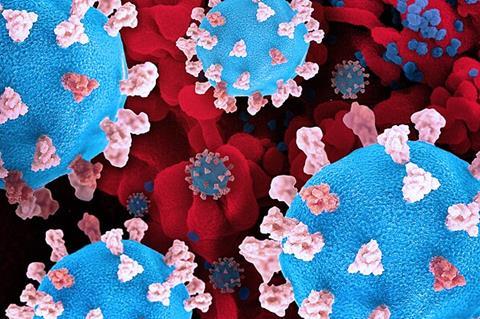
Like moths to a flame, microbes can also be moved by light. Using this knowledge, researchers from Osaka Metropolitan University’s Research Institute for Light-induced Acceleration System (RILACS) have demonstrated a method to detect the presence of viruses quickly and using only a small sample.
The research team, led by OMU Professor Takuya Iida, the director of RILACS, and Associate Professor Shiho Tokonami, the deputy director, report in npj Biosensing on a light-induced immunoassay.
READ MORE: Good as gold - improving infectious disease testing with gold nanoparticles
READ MORE: Researchers develop metal-enhanced fluorescence probes for influenza A virus detection
Using laser irradiation for less than one minute, a nanoparticle-imprinted plasmonic substrate with a series of nanobowl structures (500 nm in diameter for each) could be coated with antibodies for the spike proteins of the novel coronavirus. A 5-milliwatt laser, as low power as commercial laser pointers, could then form bubbles on the biochip that drew virus-mimicking nanoparticles, thereby accelerating the selective detection of the particles.
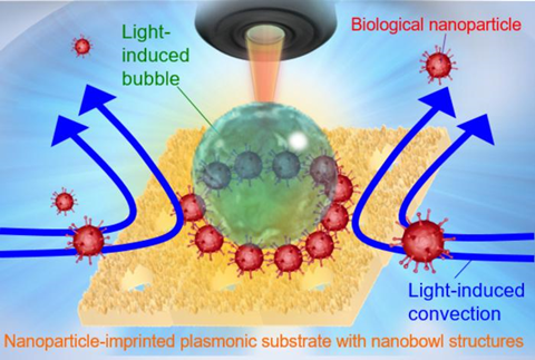
Because light-induced convection helps move the nanoparticles around so that they end up assembling at the stagnant region between the substrate surface and the bottom of the bubble, a high concentration of the particles was not required. In under five minutes, the entire process, from substrate coating to detection, could be completed.
“This study shows that we can shorten the cumbersome antibody coating process and perform rapid and highly sensitive protein detection,” Professor Iida said. “We believe our findings can contribute to the early diagnosis of not only the novel coronavirus, but possibly also various infectious diseases, cancer, even dementia.”


.jpg)
No comments yet