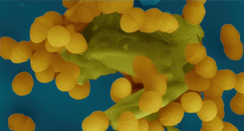
A study at Umeå University, Sweden, provides new clues in the understanding of how antibiotic resistance spreads. The study shows how an enzyme breaks down the bacteria’s protective outer layer, the cell wall, and thus facilitates the transfer of genes for resistance to antibiotics.

“You could say that we are adding a piece of the puzzle to the understanding of how antibiotic resistance spreads between bacteria,” says Ronnie Berntsson, Associate Professor at Umeå University and one of the authors behind the study.
The Umeå researchers have studied Enterococcus faecalis, which is a bacterium that often causes hospital infections, where in many cases treatment with antibiotics no longer bites because the bacteria have developed resistance. These bacteria can also spread the resistance further via the type 4 secretion systems, T4SS. It is a kind of protein complex that acts as a copying device, allowing properties in the form of genetic material to be spread to other bacteria. Resistance to antibiotics is one such trait that can be moved between bacteria with the help of T4SS.
Key enzyme
An important part of T4SS is the enzyme PrgK, which breaks down the bacterial cell wall and thus facilitates the transfer of properties between bacteria. This enzyme has three parts or domains, LytM, SLT, and CHAP.
PrgK works like scissors that cut into the bacterial cell wall. Contrary to what the researchers previously thought, it turned out that only the SLT domain was active, but in a different way than expected. The other two domains instead turned out to have an important role in the regulation of the enzyme. The researchers also identified that another T4SS protein, PrgL, binds to PrgK and ensures that it ends up in the right place in the protein machinery.
“The findings are important for continued research into how to prevent T4SS from transferring properties such as resistance to antibiotics to other bacteria,” says Josy ter Beek, Staff scientist at Umeå University.
The study has been conducted through a combination of biochemical analyses of the protein linked to functional studies in vivo, and supplemented with structural studies of PrgK using both X-ray crystallography and AlphaFold modelling.


No comments yet