Being able to observe micro-organisms and their cellular components is key to understanding fundamental processes within cells - and thus potentially developing new medical treatments.

Microbiologists and biophysicists from the University of Bonn and Wageningen University and Research have now developed a method that makes the high-throughput process for observing molecules five times faster, enabling insights to be gained into hitherto unknown cellular functions.
If our skin spends too long exposed to UV rays, e.g. from the sun, it can cause mutations in our DNA, which can potentially lead to cancer. However, the human body has a defense mechanism that it can deploy.
“Damage to our DNA activates molecules that repair it quickly, ideally before the cell divides and the damage spreads,” explains Koen Martens from the Institute for Microbiology and Biotechnology at the University of Bonn. Yet nobody quite knows exactly how fast this cellular repair function works, something that Martens now wants to find out.
Single particle tracking
This is easier said than done, however, as the methods used to date are not powerful enough to track individual molecules accurately.
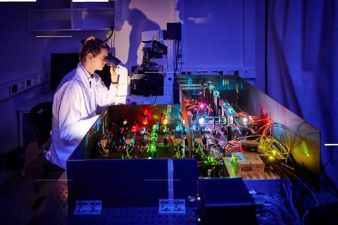
“Single particle tracking involves marking the molecule with fluorescent light, making it into a kind of light bulb,” Koen Martens explains. “We then take hundreds of photos a second using a high-resolution microscope. Our ‘light bulb’ lights up the molecule in the darkness of the cell, allowing us to observe it and track its movement over time. This enables us to measure its diffusion and how it interacts with other cellular components.”
By looking at the gaps between molecules and the distances traveled by a single molecule from one photograph to another, the researchers can tell whether the particles are moving freely inside the cell or interacting with other molecules. As far as DNA repair is concerned, this indicates when the enzymes are performing their repair work—i.e. when they are interacting with the DNA—and when they are “idle,” i.e. diffusing freely inside the cell.
One drawback
However, the method does have one drawback: “It’s hard to track multiple molecules at the same time,” Martens explains. “When their paths cross or they’re too close together, you get two light bulbs merging, in effect. Then it’s impossible to identify their movements.”
Up until now, therefore, the microbiologists have had to study molecules one after the other in a time-consuming process that is too long-winded to observe the DNA-repairing molecules “at work.” In fact, single particle tracking currently takes longer than the repair process itself.
To solve the problem, Koen Martens has created a piece of software to speed up the high-throughput process. TARDIS (short for “temporal analysis of relative distances”) runs an all-to-all analysis of the distances between locations, i.e. the positions of the molecule in the individual photographs, with increasing time lags. Instead of focusing on individual points as before, it looks at the entire sequence of movements within the cell and thus scrutinizes all the molecules simultaneously. “TARDIS makes the measurement process at least five times faster without any loss of information,” says Martens.
Next step
This means that he can now devote his attention to the remaining part of his research project, using TARDIS to study the processes involved in DNA repair in more detail. “I’m especially interested in investigating how easy or difficult certain kinds of damage are to repair and how badly the DNA is damaged by a specific dose of UV radiation or chemicals.
Wageningen University & Research was involved in this study alongside the University of Bonn. The work was funded by the Alexander von Humboldt Foundation, Carnegie Mellon University, the Argelander Program for Early-Career Researchers at the University of Bonn, the PhD Fellowship at the VLAG Graduate School of Wageningen University, and the National Science Foundation.


No comments yet